The ABCs of Cranial Cruciate Ligament Disease

What to look for in your radiographic study


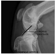
  • The cranial cruciate ligament goes from the medial surface of the lateral femoral condyle to the cranial proximal surface of the tibia.
  • The ligament stabilizes the stifle against cranial-caudal and internal rotation of the tibia.
  • Most clinicians evaluate the integrity of this ligament by palpating for abnormal motion.
  • In cases where the ligament is not completely torn or where fibrosis has stabilized the joint, laxity may not be present-the author states this occurs 40% of the time in the cases he sees.
  • The radiographic findings of cruciate ligament disease then become paramount in
  • detecting this condition.
  • Other imaging methods-arthroscopy/MRI can be used to assess the stifle joint;  however, the survey radiograph study continues to be the first method of imaging assessment in most veterinary practices.
abcs normalstifle
Normal Stifle


No evidence of joint effusion


No evidence of bone reaction where the cranial cruciate inserts into the proximal cranial surface of the tibia.
abcs abnormalstifle
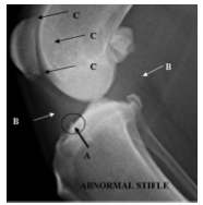
Abnormal Stifle


A. Osseous reaction where cruciate inserts into the tibia.

B. Increased soft tissue volume in the joint.

C. Multicentric areas of new bone production and loss.


Even if there is no joint laxity, if there is reaction where the cruciate inserts into the proximal tibia, then the cruciate ligament is involved in the disease process.